

AI-Powered 3D Segmentation for Cardiac Imaging
NeuralFlex developed AI-powered 3D medical image segmentation technology for detailed anatomical reconstruction and analysis. The work contributed to cardiac imaging research published in Nature Cardiovascular Research, demonstrating the use of deep-learning-based segmentation within an advanced 3D visualization workflow.
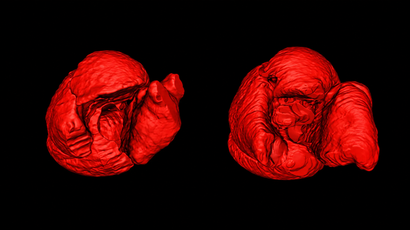
01 / OVERVIEW
Transforming cardiac imaging into detailed 3D structural intelligence.
Cardiac imaging contains complex anatomical structures that can be difficult and time-consuming to isolate manually across volumetric scan data.
NeuralFlex developed a deep-learning-based segmentation workflow designed to identify cardiac structures from CT imaging and transform them into detailed three-dimensional reconstructions.
By automating the segmentation process and connecting imaging data with 3D visualization, the solution provides a foundation for more detailed structural analysis and advanced cardiac imaging research.
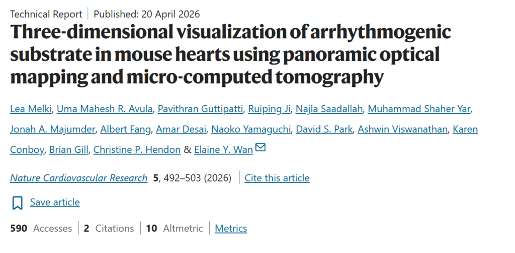
Published in Nature Cardiovascular Research
This work contributed to peer-reviewed cardiac imaging research combining micro-computed tomography, three-dimensional visualization, and deep-learning-based segmentation to support detailed analysis of cardiac structures.
Nature Cardiovascular Research
Technical Report · Published 20 April 2026
See cardiac segmentation and 3D Reconstruction in action.
Explore the segmentation workflow from cardiac CT imaging and predicted anatomical masks to detailed three-dimensional reconstruction.
Segmentation Analysis
Cardiac CT imaging with segmentation masks and predicted anatomical regions.
3D Cardiac Reconstruction
Three-dimensional visualization of the segmented cardiac anatomy from multiple perspectives.
02 / THE CHALLENGE
Cardiac anatomy is complex to isolate across volumetric imaging data.
Cardiac CT data contains complex anatomical boundaries across hundreds of image slices, making consistent segmentation a challenging and time-intensive task.
Manual delineation requires structures to be identified across multiple views while maintaining anatomical consistency throughout the volume. This becomes particularly demanding when the segmented data must later support accurate three-dimensional reconstruction and structural analysis.
The challenge was to develop an automated approach capable of identifying cardiac structures across volumetric imaging data and producing segmentation outputs suitable for detailed 3D visualization.
NeuralFlex developed a deep-learning segmentation pipeline that processes volumetric cardiac imaging and identifies anatomical regions across individual scan slices.
The predicted segmentation masks are combined across the imaging volume to reconstruct the segmented cardiac structures in three dimensions. This creates a continuous representation of the anatomy that can be inspected from different perspectives and used for further structural analysis.
The workflow connects medical image processing, automated segmentation, and 3D reconstruction within a unified cardiac imaging pipeline.
03 / THE Architecture
From volumetric CT data to reconstructed cardiac anatomy.
04 / WHAT NEURALFLEX BUILT
Engineering an automated cardiac segmentation workflow.
01
Cardiac Image Segmentation
Processes volumetric CT imaging to identify and isolate relevant cardiac structures across individual scan slices.
02
Deep-Learning Segmentation Pipeline
Uses deep-learning-based image segmentation to generate anatomical masks and automate a process that would otherwise require extensive manual delineation.
03
3D Cardiac Reconstruction
Combines segmented regions across the imaging volume to generate detailed three-dimensional representations of cardiac anatomy.
04
Visualization & Structural Analysis
Enables reconstructed cardiac structures to be inspected from multiple perspectives, providing a foundation for detailed anatomical and structural analysis.
05 / RESEARCH APPLICATIONS
Supporting advanced cardiac imaging and structural research.
The segmentation system is designed to support medical professionals by transforming complex imaging data into clearer anatomical information for analysis and planning.
01 / CARDIAC IMAGING
Converts volumetric cardiac scans into structured anatomical segmentations for detailed visualization and analysis.
02 / 3D ANATOMICAL ANALYSIS
Reconstructs segmented cardiac structures in three dimensions, enabling researchers to examine anatomy from multiple perspectives.
03 / COMPUTATIONAL RESEARCH
Provides structured imaging outputs that can support advanced computational analysis and cardiac research workflows.
06 / THE RESULT
From complex cardiac scans to structured 3D anatomical insight.
The result is an AI-powered imaging workflow that transforms volumetric cardiac CT data into segmented anatomical structures and detailed three-dimensional reconstructions.
By combining deep-learning-based segmentation with 3D visualization, NeuralFlex reduced the manual effort involved in extracting cardiac structures while creating consistent outputs for structural analysis and research workflows.
The technology contributed to peer-reviewed cardiac imaging research published in Nature Cardiovascular Research, demonstrating how AI-based segmentation can form part of advanced three-dimensional cardiac visualization workflows.